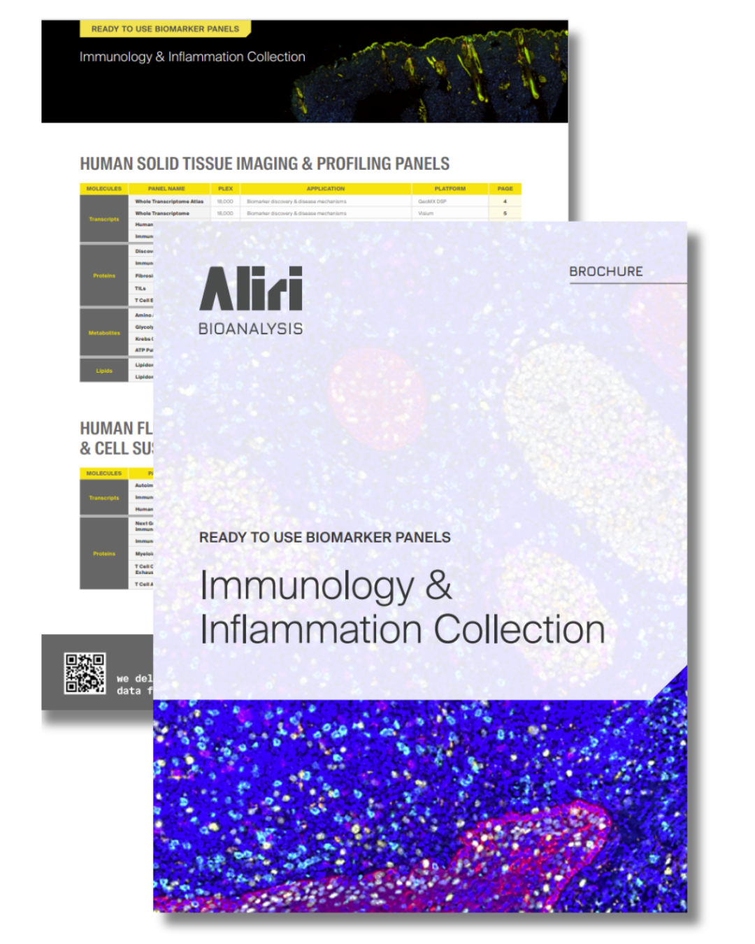
Ready-to-Use Biomarker Panels: Immunology & Inflammation Collection
From disease onset to therapeutic response, Immunology and Inflammation research increasingly relies on biomarker data that must be robust, translatable, and biologically meaningful. Clinicians and translational teams now require the simultaneous assessment of growing numbers of biomarkers to better capture disease complexity, patient heterogeneity, and treatment response.
To expedite your research and minimize development costs, we have translated our years of immunology and inflammatory biomarker expertise into 23 ready-to-use panels. These panels are designed to address key disease-relevant questions about disease onset, progression, and response to therapy, including for example:
- Immune cell activation and differentiation
- Cytokine and chemokine signaling
- Tissue inflammation
- Pathway dysregulation
- Patient stratification
Download the brochure to view a full list of panels and biomarkers in our Immunology & Inflammation Collection.
ABCs of ADCs: From PK to PD

Predictive bioanalytical strategies for the development of Antibody-Drug Conjugates (ADCs)
ADCs often fail because the drug gets to the right tissue, but misses its target. It’s not enough to just seek the concentration of your ADC in the tissue, you need to know its exact location to predict efficacy and move your development program forward with confidence. In this webinar, renowned scientists and industry thought leaders Troy Voelker and Corinne Ramos outline a holistic bioanalytical strategy for your ADC, incorporating traditional bioanalysis with spatial bioanalysis to reveal decision-making data about how your ADC engages with the complex microenvironment.
- Antibody payload concentration
- Conjugated antibody concentration
- Free payload concentration
- Spatial distribution
- Target engagement
- Toxicity
The Complexity of the Gastrointestinal Tract: A Multilayered Challenge for Drug and Biomarker Studies
The gastrointestinal (GI) tract is a highly specialized and dynamic system, playing a central role in digestion, absorption, immune response, and microbiome interaction. From the stomach to the small intestine (duodenum, jejunum, ileum) and the large intestine (colon, rectum), each segment presents unique physiological and structural characteristics that influence drug delivery and disease progression.
Aliri’s spatial imaging capabilities allows you to understand drug behavior and biomarker expression across the four main layers of the intestinal wall.
Download the fact sheet to learn more.
Oligonucleotide Hybridization LCMS Workflows and Probe Optimization
In this scientific poster recently presented at EBF Open Symposium, we investigated the biodistribution and potential toxicity of lipid nanoparticles (LNP1 and LPN2), which are crucial carriers for mRNA-based treatments after administration to male and female mice, analyzing their distribution in whole-body carcasses and specific organs using MALDI-MSI.
Download our poster to learn more.
Mapping mRNA–Lipid Nanoparticle Distribution in Mouse Whole Body and Organs by MALDI-MSI
In this scientific poster recently presented at EBF Open Symposium, we investigated the biodistribution and potential toxicity of lipid nanoparticles (LNP1 and LPN2), which are crucial carriers for mRNA-based treatments after administration to male and female mice, analyzing their distribution in whole-body carcasses and specific organs using MALDI-MSI.
Download our poster to learn more.
Ring Trial Study Results for Oligonucleotides Prove Hybridization LC-MS Approach Superior to LBA in Achieving Lower Detection Limits and Higher Specificity
Aliri was recently one of 10 labs to participate in the Oligonucleotide Ring Trial, in which industry experts joined together to evaluate the effectiveness of LC-MS, LBA, and qPCR when quantifying the concentrations of oligonucleotide in biological samples. This first-of-its-kind study aimed to give clarity to drug developers about the methodology best suited for future development programs. Specifically, the Ring Trial focused on three types of oligonucleotides, an ASO (Fomivirsen), a GalNAc-siRNA (Lumasiran), and a PMO (Viltolarsen).
Troy Voelker, Sr. Lab Director at Aliri and Chair of the AAPS Oligonucleotide Discussion Group, led the LC-MS method development of the PMO (Viltolarsen), which was analyzed using three mass spectrometry platforms: a QExactive, a time-of-flight (TOF), and a triple quadrupole instrument. In this presentation, he reveals exciting data that proves hybridization LC-MS superior to LBA in achieving lower detection limits and higher specificity.
Download the presentation to read the study findings.
Enabling Clinical Adoption of Omics: Fit-for-Purpose Validation and a Spatial Biomarker Case Study
Over the past two decades, omics technologies have steadily expanded from discovery research into translational and clinical development, offering unprecedented insights into biology, disease mechanisms, and therapeutic response. Advances in genomics, transcriptomics, proteomics, metabolomics, and, more recently, spatial and multi-omics platforms have created powerful opportunities to identify predictive biomarkers, refine patient stratification, and accelerate drug development. The added value of omics lies in their ability to capture complex, system-level biology that traditional single-analyte assays cannot address, thereby bridging the gap between exploratory research and precision medicine.
Yet this rapid progress has also highlighted a critical challenge: how to validate omics data in a way that is scientifically rigorous but also practical. Current analytical validation paradigms were largely developed for conventional assays such as ELISA or qPCR, which measure one or a handful of targets at a time. Applying the same frameworks directly to high-dimensional omics assays often results in processes that are overly burdensome, expensive, and poorly aligned with the dynamic nature of omics platforms.
Moving forward, the field needs more efficient and adaptive validation processes, aligned with the specific purpose of each omics application. This involves applying full rigor when data will inform regulatory submissions or clinical decisions, while using streamlined, fit-for-purpose approaches for exploratory research and mechanistic studies.
In this presentation, Aliri R&D Director and Spatial-Omics Expert, Corinne Ramos, Ph.D., illustrates these challenges and opportunities through a spatial multi-omics use case, where paired patient biopsies were profiled with spatial transcriptomics and proteomics to uncover mechanistic insights, identify predictive biomarkers, and generate regulatory-ready evidence for clinical development.
Download the presentation to learn more.
Maximizing Reproducibility and Sensitivity in qPCR for Detecting Transcripts Over a Broad Dynamic Range in Response to Anti-PD-1 Therapy
This study aims to optimize and validate a qPCR workflow for the reproducible and sensitive quantification of immune checkpoint transcripts (PD-1,PD-L1, CTLA-4) in FFPE lung cancer tissues. By refining tissue preparation, RNA input, and assay conditions, we establish a robust method for detecting gene expression across a broad dynamic range, enabling reliable assessment of immunotherapy response and supporting biomarker-driven patient stratification.
Download our poster to learn more.
Development and Validation of a Sensitive LC-MS/MS Method for the Quantification of SGR-1505 in Human Plasma to Support Clinical Pharmacokinetic Studies
MALT1 is a key mediator of NF-κB signaling and an emerging therapeutic target in B-cell malignancies and autoimmune diseases. SGR-1505, a potent MALT1 inhibitor, is being clinically evaluated for its therapeutic potential. In this study, we developed and validated a reliable and high-throughput LC-MS/MS method for the quantitation of SGR-1505 in human plasma (K₂EDTA) to support clinical pharmacokinetic studies. This work exemplifies the critical role of CRO-led bioanalysis in bridging early discovery and clinical development of emerging therapeutics.
Download our poster to learn more.
Development of Total ASO method in Mouse Plasma and Tissues Using LC-FD and LC-MS Platforms
Bioanalytical methods are needed to analyze protein conjugated antisense oligonucleotides (POCs) to accurately quantify the active antisense oligonucleotide (ASO) payloads, assess its pharmacokinetics and biodistribution in plasma and tissues, and ensure patient safety by evaluating potential immunogenicity and toxicity. Because the conjugate, the free ASO, and the linked ASO fragment can all be present, specialized techniques are required to differentiate and quantify these components, which is essential for supporting the development of these complex biotherapeutics. The unique properties of POCs present significant analytical challenges that necessitate specialized methods.
We set out to develop a methodology for quantifying total ASO in POCs that could be universally applied across similar POCs. The
study compared mass spectrometry and fluorescence detection platforms to determine optimal sensitivity, selectivity,
and adaptability. Additionally, it aimed to establish a single extraction method suitable for both plasma and tissue
analysis.
Download our poster to learn more.