Single Clinical Trial Strategy to Accelerate your Drug’s Path to Market
Recent FDA discussions surrounding plausible mechanism and confirmatory evidence frameworks reflect this broader shift. Increasingly, translational success may depend not only on demonstrating that a therapy produced a measurable effect, but also on confirming how that effect emerged within intact biological systems. FDA guidance and recent regulatory discussions increasingly reference mechanistic, pharmacodynamic, and target engagement evidence as important components of confirmatory evidence frameworks.
As spatial biology moves closer to regulated translational and clinical development environments, the challenge is no longer simply generating more molecular data. The next phase of the field will depend on the ability to generate biologically coherent, reproducible, scalable, and clinically actionable mechanistic evidence.
Download the white paper to learn how spatial biology can support the FDA’s Evolving Confirmatory Evidence Framework.
A Spatial PK/PD Framework to Predict ADC Response in Solid Tumors
Antibody-drug conjugates achieve therapeutic activity only when payload delivery, target accessibility, and microenvironmental readiness align within tumor tissue. Conventional biomarkers capture isolated components of this biology but fail to resolve the spatial coordination that ultimately governs pharmacodynamic activity.
To address this gap, we developed a translational spatial PK/PD framework integrating MSI, spatial proteomics/transcriptomics, and multiplex tissue imaging to spatially interpret coordinated ADC activity within solid tumors
Download the poster, recently presented at EACR 2026 in Budapest, Hungary to learn more.
Qualification of Translational Spatial Biomarker Panel to Enable Predictive Tissue Profiling in Oncology Drug Development
Spatial biology technologies are transforming translational oncology by enabling high-resolution characterization of tumor–immune ecosystems. However, the clinical utility of spatial biomarkers remains limited by a lack of standardized, qualified panels capable of generating reproducible and decision-enabling data across studies. To address this gap, we developed a framework for the qualification of translational spatial biomarker panels designed to support drug development and clinical trial biomarker strategies. By integrating multiplex immunofluorescence, digital pathology, and AI-assisted image analysis, our objective was to establish reproducible workflows capable of generating biologically relevant and analytically robust spatial biomarkers suitable for translational oncology applications.
Download the poster, recently presented at EACR 2026 in Budapest, Hungary to learn more.
Spatial Multi-Omics Meets AI: Turning Tissue into Actionable Insight, Designing Insightful Spatial Multi-Omics Studies: From Sample to Signal
Precision medicine requires technologies capable of capturing both molecular heterogeneity and tissue architecture. Unlike conventional bulk approaches, spatial multi-omics preserves tissue context, enabling the characterization of genes, proteins, metabolites, and cell–cell interactions within their native environment. Combined with AI-driven image analysis and multimodal data integration, spatial multi-omics provides a powerful framework for biomarker discovery, patient stratification, and treatment response prediction.
To demonstrate the potential of AI-enabled spatial multi-omics for precision medicine, we applied an established machine learning framework to identify treatment-associated molecular signatures in cervical cancer using integrated proteomic, transcriptomic, and metabolomic data.
Download our poster, recently presented at AI in Oncology Paris to learn more.
Aliri Clinical APEX Process Overview
Aliri’s Clinical Analysis and Precision Execution (APEX) Process is built to run large clinical trials without the loss of scientific control, ensuring data remains robust, interpretable, and decision-ready as studies grow.
It’s not just about capacity. Clinical studies succeed or fail in the details. Selecting a strong CRO partner for your molecule’s late-phase bioanalysis means prioritizing:
- Direct access to senior scientists
- Industry-leading, fit-for-purpose methods
- Embedded automation
- The best-available instruments for the job
- Regulatory-ready, defensible data
- A team you never have to wait on
Download the fact sheet to learn more.
Case Study: Actionable, Qualified Spatial Profiling of Pharmacodynamic-Relevant Tumor–Immune Biology in NSCLC
The next evolution of spatial biology is the transition from exploratory imaging toward controlled generation of quantitative spatial metrics associated with pharmacodynamic-relevant tumor biology.
In non-small cell lung cancer (NSCLC) immuno-oncology studies, the challenge is not simply identifying immune cells within the tissue, but understanding whether their spatial organization reflects biologically meaningful mechanisms associated with therapeutic activity, immune exclusion, suppression, or cytotoxic engagement.
This case study illustrates how a qualified multiplex spatial panel can be deployed on FFPE NSCLC tissue to generate reproducible and biologically interpretable spatial metrics associated with pharmacodynamic (PD)-relevant tumor–immune biology. The objective: implementation of a controlled multiplex framework capable of moving beyond descriptive multiplex imaging toward more quantitative spatial characterization of immune biology.
Download the case study below.
Ready-to-Use Standalone Biomarkers
Understanding tissue biology at high resolution requires both flexibility and reliability. At Aliri, we provide a curated portfolio of verified standalone protein markers compatible with immunofluorescence and imaging mass cytometry (IMC) workflows.
These markers are designed to seamlessly complement our ready-to-use panels or to be deployed as individual targets, enabling tailored experimental designs without compromising on quality or timelines.
Download the full list of standalone biomarkers below.
Optimizing Surrogate Matrix Selection for Endogenous Biomarker LC-MS/MS Quantitation Assays
Historically, LC-MS/MS focused on synthetic drug pharmacokinetics. However, improved sensitivity now allows the analysis of endogenous biomarkers (lipids, proteins, etc.) previously reserved for Ligand Binding Assays (LBA). A primary challenge in biomarker bioanalysis is that these compounds are endogenous to biological matrices, and the matrix could present challenging physicochemical properties. Unlike conventional drug testing, there is no true blank matrix available to build calibration standards. To quantify these levels accurately, researchers must develop a surrogate matrix that mimics the behavior of authentic patient samples without the background interference of the natural analyte. Selecting and validating this surrogate is the most critical step in establishing a reliable LC-MS/MS method for any naturally occurring compound.
Download our poster, recently presented at WRIB 2026 to learn more.
Advanced Bioanalytical Support for Oligonucleotides
Aliri has supported 5 of the 20 approved OGNs on market as they transitioned from preclinical to clinical to FDA approval Aliri is proud to be a trailblazer in the bioanalysis of RNAi therapeutics with significant experience supporting the development of oligonucleotides, which includes siRNAs, ASOs, PPMOs, ARCs/AOCs, and other RNAi conjugates.
Our tenured team of scientists are continuously innovating and adapting our methodologies for improved
assay sensitivity and specificity to deliver accurate and reliable data to our sponsors.
This has led to the critical application and optimization of available analytical approaches, including novel SPE and hybridization extraction techniques that can achieve subnanogram per milliter LLOQ’s with the
specificity of LC/MS detection.
Download the fact sheet to learn more.
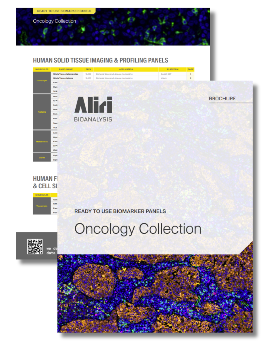
Ready-to-Use Biomarker Panels: Oncology Collection
From tumor initiation to therapeutic response, oncology research increasingly depends on biomarker data that must be robust, translatable, and clinically meaningful. Clinicians and translational teams now require the simultaneous assessment of an expanding number of biomarkers to better capture tumor heterogeneity, mechanisms of resistance, and patient response to therapy.
To expedite your research and minimize development costs, we have translated our years of oncology biomarker expertise into 24 ready-to-use panels. These panels are designed to address key oncology questions that are critical for understanding disease biology and therapeutic response, including for example:
- Tumor characterization
- Immune contexture
- Pathway activation
- Target engagement
- Patient Stratification
Download the brochure to view a full list of panels and biomarkers in our Oncology Collection.