The Hyperion™ Imaging System is an advanced high-plex immuno-staining platform that leverages Imaging Mass Cytometry (IMC) to provide high-resolution, multiplexed spatial proteomics data. Developed by Fluidigm (now part of Standard BioTools), the Hyperion™ Imaging Mass Cytometry utilizes antibodies conjugated to stable metal isotopes instead of traditional fluorophores or chromogens. These metal-tagged antibodies bind to specific proteins within the tissue sample, and the laser ablation system scans the tissue, releasing the bound isotopes. These isotopes are then detected by a time-of-flight mass spectrometer (CyTOF), generating a detailed spatial proteomic map of the tissue.
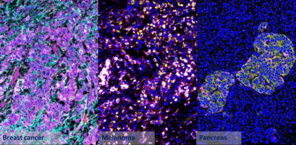
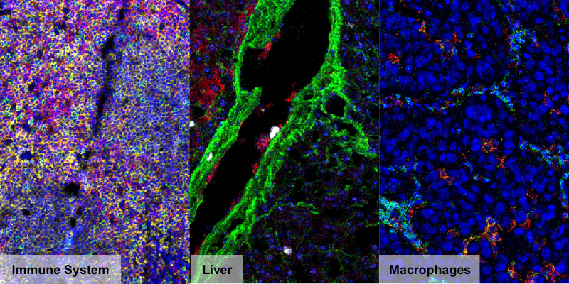
The Hyperion™ Imaging System revolutionizes spatial proteomics by enabling the simultaneous visualization of 40+ biomarkers in a single tissue section at subcellular resolution (1 μm). Unlike traditional fluorescence-based multiplexing methods, Hyperion™ avoids spectral overlap and autofluorescence interference, ensuring clear, quantitative, and highly reproducible data. By utilizing metal-tagged antibodies and laser ablation coupled with time-of-flight mass spectrometry (CyTOF), this approach provides deep tissue penetration, absolute quantification, and detailed spatial mapping of the tumor microenvironment (TME), immune infiltration, and cellular interactions. Combined with Aliri’s powerful proprietary image analysis, this innovative capability helps our clients evaluate targeted biomarkers for preclinical and clinical samples and classify patients based on tissue biomarkers.